Describe the Anatomy of a Sponge
Sponges have special collar cells or choanocytes that are unique in the animal kingdom. Based on your explanations what would you expect to be thicker and heavier.
Anatomy The Basics Of Porifera
The marine worm Ramisyllis multicaudata which lives within the internal canals of a sponge is one of only two such species possessing a branching body with one head and multiple posterior ends.

. Figure 631 Anatomy of a Long Bone. See the answer Describe the anatomy of a sponge. They have flagella whip-like structures that work to set up water currents so the sponge can sieve food particles.
As the most ancient extant metazoans glass sponges hexactinellida have attracted recent attention in the areas of molecular evolution and the evolution of conduction systems but they are also interesting because of their unique histology. They obtain food and oxygen and remove waste through this water movement. Compressed polygonal cells called pinacocytes make up the pinacoderm the external sac layer.
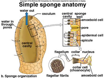
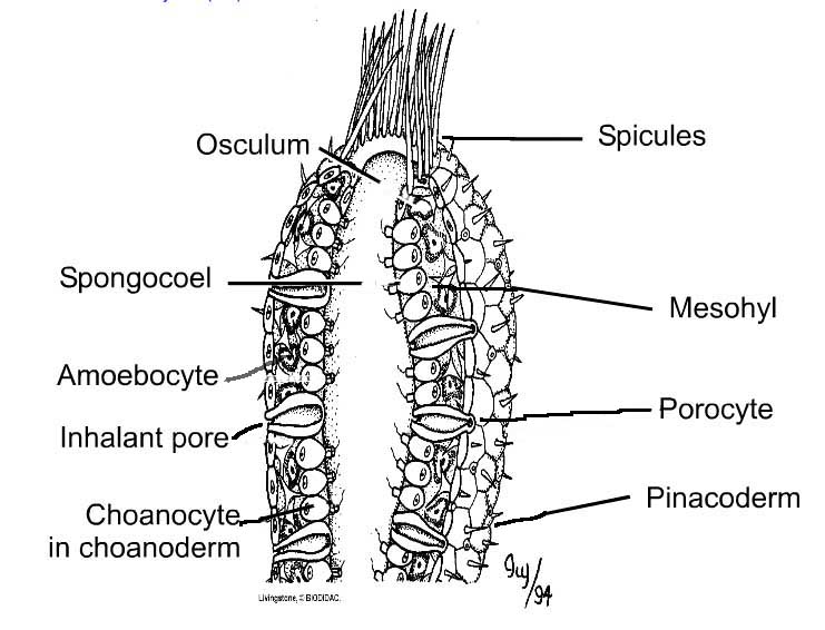
These pores have given the sponges their phylum name Poriferapore-bearers. The marine worm Ramisyllis multicaudata which lives within the internal canals of a sponge is one of only two such species possessing a branching body with one head and multiple posterior ends. Sponges are animals with dense skeletons that are highly adapted to their environments although it is easy to see why they may be mistaken for plants.
Describe the anatomy and life cycle of sponges andcnidarians. Describe and draw the basic anatomy of a sponge including. Be sure to include the names of the major types of cells and their functions.
Red bone marrow fills the spaces between the spongy bone in some long bones. Simple sponges with choanocytes lining inner cavity radially symmetrical Syconoid Condition Choanoderm and Pinacoderm folding creates the shape. Sponges have neither tissues nor organs.
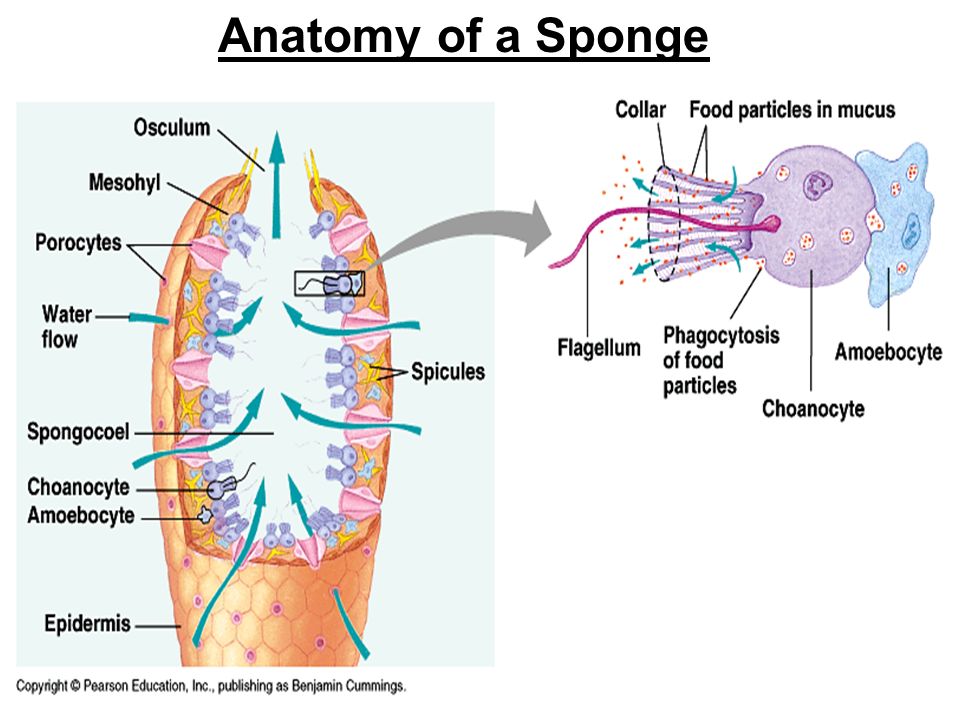
Since sponges do not have real tissues or organs therefore they have no digestive system such as that of more complex living things their only. In asconoid sponges the two major cell layers surround a fluid-filled cavity called the spongocoel the large central cavity of sponges. Sponges belong to phylum Porifera and are asymmetrical sessile and non-moving.
Scattered among the pinacoderm are the ostia that allow entry of water into the body of the sponge. Instead they consist of three cell sized layers. Sponges are found in a wide variety of colors shapes and sizes and scientists believe that the colors of the sponge may act as a protection from the suns harmful UV rays.
The greater part of their soft tissue consists of a single multinucleate syncytium that ramifies. Sponges are exclusively aquatic filter feeders that actively pump water through their bodies to eat breathe and excrete. The morphology of the simplest sponges takes the shape of a cylinder with a large central cavity the spongocoel occupying the inside of the cylinder.
Water can enter into the spongocoel from numerous pores in the body wall. Ostia oscula choanocytes amoebocytes spicules Ostia small incurrent pores Oscula large excurrent pores Choanocytes collar cells filter plankton Spicules skeleton-like glass or calcium Amoebocytes - Secrete spicules - Produce gametes sperm eggs. Water entering the spongocoel is extruded via a large common opening called the osculum.
It consists of a cell mass in the form of a sac through which the water circulates in which is the oxygen that allows it to breathe and the food with which it subsists. In the gel layer are either spicules supportive needles made of calcium carbonate or spongin fibers a flexible skeletal material made from protein. The chambers scattered throughout the body of the sponge have pores through which water passes into a complex system of incurrent canals then into a spongocoel internal cavity by way of excurrent canals.
The cells in the outer. A typical long bone showing gross anatomical features. The body of a sponge has two outer layers separated by an acellular having no cells gel layer called the mesohyl also called the mesenchyme.
The sponge is made up of two single-cell-deep layers and an intermediate mesohyl mobile cells plus extracellular matrix. In male anatomy the corpus. Water enters very small pores found among the cells pinacocytes which line the outer surface of the sponge.
The three groups of tissues the two cavernosa and the spongiosum are expandable sponge-like structures. Best Answer 100 1 rating The body of a sponge has two outer layers separated by an acellular having no cells gel layer called. Contrastingly what effect does sedentarism weightlessness in space or in bedridden patients.
The wider section at each end of the bone is called the epiphysis plural epiphyses which is filled internally with spongy bone another type of osseous tissue. Up to 24 cash back Basic Anatomy. The middle mesohyl layer consists of gelatinous proteincarbohydrate material a range of mobile cells and a skeleton of calcareous or siliceous spicules or of elastic.
They have a unique feeding system in which they keep constant water flow through their bodies. Choanocytes in small chambers called choanocyte chambers original much that open to an apopyle leading to the spongocoel. Sponges contain no organs or even tissue.
The outer sac layer consists of flattened polygonal cells called pinacocytes. One of the main digestive cell types is the choanocyte. The collar cells of sponges trap and digest food.
View the full answer Previous question Next question. In some sponges ostia are formed by porocytes single tube-shaped cells that act as valves to regulate the flow of water into the spongocoel. Sponges are multi-cellular organisms which are devoid of organ systems like digestive system nervous system circulatory system et cetera.
The arm bones of a weightlifter or the arm bones of a jogger. Describe the specific changes seen in the microscopic anatomy of the sponge bone in response to exercise. Feeding Sponges Responds to its anatomical structure which is quite simple.
An international research team led by the Universities of Göttingen and Madrid is the first to describe the internal anatomy of this intriguing. This problem has been solved. A sponge lacks tissues and organs but it has several types of specialized cells.
Water is pumped directly through pores called ostia into the spongocoel and then out of the sponge through an opening called the osculum plural oscula.
Review Of Animal Phylogeny Sponges Anatomy Of A Sponge Ppt Download
Overview Of Sponges
Sponge Structure And Function Advanced Read Biology Ck 12 Foundation
No comments for "Describe the Anatomy of a Sponge"
Post a Comment